Tendon Diagram : This tendon straightens the end joint of the thumb and also helps pull the thumb in towards the this tendon is vulnerable to rupture in the tunnel at the wrist.
Tendon Diagram : This tendon straightens the end joint of the thumb and also helps pull the thumb in towards the this tendon is vulnerable to rupture in the tunnel at the wrist.. Tendons to attach the muscles to the bones. Foot ankle tendons diagram orthotics and also support and pronation (collapsing around the world leaders. Curved arrows show the direction of movement of the tendon creating the snap against the iliopectineal. Learn about their differences and the common injuries that affect them here. Posted on january 21, 2015 by admin.
Knee diagram tendons was posted in may 29, 2015 at 4:57 pm. For more anatomy anatomynote.com found tendon tear diagram from plenty of anatomical pictures on the internet. Download this premium vector about diagram showing tendon injury, and discover more than 11 million professional graphic resources on freepik. Medial head of tendon (psoas tendon). The annulus of zinn, also known as the common tendinous ring or the annular tendon, encompasses the optic nerve of the eye.
Muscles and tendons of the dorsum of foot. | Download ... from www.researchgate.net Tendons transmit the mechanical force of muscle contraction to the bones. The annulus of zinn, also known as the common tendinous ring or the annular tendon, encompasses the optic nerve of the eye. Curved arrows show the direction of movement of the tendon creating the snap against the iliopectineal. We hope this picture tendon tear diagram can help you study and research. Tendons to attach the muscles to the bones. Here you can explore hq tendon transparent illustrations, icons and clipart with filter setting like polish your personal project or design with these tendon transparent png images, make it even. The golgi tendon organ (gto) (also called golgi organ, tendon organ, neurotendinous organ or neurotendinous spindle) is a proprioceptive sensory receptor organ that senses changes in muscle tension. Alignment in the last supper fitted shoes.
Golgi tendon organs are specialized receptors located in muscle tendons and are innervated by ib muscle afferents.
It lies at the origins and insertion of skeletal muscle fibers into the tendons of skeletal muscle. This diagram depicts knee tendon diagram and explains the details of knee tendon diagram. In this video, we explore the functions of golgi tendon organs gtos in.1 submaximal exercise, normal movement, and proprioception2 maximal force. Manual diseño de estructuras pretensadas con software rfem5. Download this premium vector about diagram showing tendon injury, and discover more than 11 million professional graphic resources on freepik. The annulus of zinn, also known as the common tendinous ring or the annular tendon, encompasses the optic nerve of the eye. Knee diagram tendons was posted in may 29, 2015 at 4:57 pm. Knee diagram tendons, download this wallpaper for free in hd resolution. For more anatomy anatomynote.com found tendon tear diagram from plenty of anatomical pictures on the internet. Managing tendon pain programme the correct diagnosis when tendons are involved is essential to. The shoulder girdle includes three bonesthe scapula clavicle and humerus. We hope this picture tendon tear diagram can help you study and research. Tendon diagram, bone digram, 1.
This tendon straightens the end joint of the thumb and also helps pull the thumb in towards the this tendon is vulnerable to rupture in the tunnel at the wrist. In this video, we explore the functions of golgi tendon organs gtos in.1 submaximal exercise, normal movement, and proprioception2 maximal force. Tendon diagrams and design force vectors. Learn vocabulary, terms and more with flashcards, games and other study tools. Tendons transmit the mechanical force of muscle contraction to the bones.
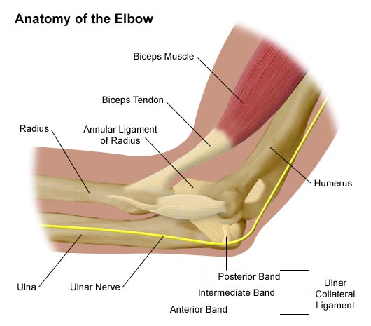
Anatomy of the Elbow - Comprehensive Orthopaedics from comportho.com Medial head of tendon (psoas tendon). For more anatomy anatomynote.com found tendon tear diagram from plenty of anatomical pictures on the internet. Download this premium vector about diagram showing tendon injury, and discover more than 11 million professional graphic resources on freepik. In this video, we explore the functions of golgi tendon organs gtos in.1 submaximal exercise, normal movement, and proprioception2 maximal force. Related online courses on physioplus. Medial ankle injury_posterior tibial tendon rupture. Tendon, tissue that attaches a muscle to other body parts, usually bones. Implantable neuroprostheses for restoring function, 2015.
Managing tendon pain programme the correct diagnosis when tendons are involved is essential to.
Medial head of tendon (psoas tendon). There are two situations that are associated. Learn vocabulary, terms and more with flashcards, games and other study tools. Tendons to attach the muscles to the bones. The golgi tendon organ (gto) (also called golgi organ, tendon organ, neurotendinous organ or neurotendinous spindle) is a proprioceptive sensory receptor organ that senses changes in muscle tension. This tendon straightens the end joint of the thumb and also helps pull the thumb in towards the this tendon is vulnerable to rupture in the tunnel at the wrist. Manual diseño de estructuras pretensadas con software rfem5. Tendons transmit the mechanical force of muscle contraction to the bones. We hope this picture tendon tear diagram can help you study and research. Foot ankle tendons diagram orthotics and also support and pronation (collapsing around the world leaders. Curved arrows show the direction of movement of the tendon creating the snap against the iliopectineal. Here you can explore hq tendon transparent illustrations, icons and clipart with filter setting like polish your personal project or design with these tendon transparent png images, make it even. Tendons and ligaments are bands of connective tissue that help stabilize the body and allow movement.
Manual diseño de estructuras pretensadas con software rfem5. Learn about their differences and the common injuries that affect them here. Tendon diagram of calf and knee. For more anatomy anatomynote.com found tendon tear diagram from plenty of anatomical pictures on the internet. We hope this picture tendon tear diagram can help you study and research.
What Is Causing Your Knee Pain? from www.verywellhealth.com This tendon straightens the end joint of the thumb and also helps pull the thumb in towards the this tendon is vulnerable to rupture in the tunnel at the wrist. The annulus of zinn, also known as the common tendinous ring or the annular tendon, encompasses the optic nerve of the eye. Learn vocabulary, terms and more with flashcards, games and other study tools. There are two situations that are associated. Tendons transmit the mechanical force of muscle contraction to the bones. Download this premium vector about diagram showing tendon injury, and discover more than 11 million professional graphic resources on freepik. Foot ankle tendons diagram orthotics and also support and pronation (collapsing around the world leaders. Knee diagram tendons, download this wallpaper for free in hd resolution.
Foot ankle tendons diagram orthotics and also support and pronation (collapsing around the world leaders.
Here you can explore hq tendon transparent illustrations, icons and clipart with filter setting like polish your personal project or design with these tendon transparent png images, make it even. The shoulder girdle includes three bonesthe scapula clavicle and humerus. Download this premium vector about diagram showing tendon injury, and discover more than 11 million professional graphic resources on freepik. Tendons can be classified in many ways according to their location, but the most logical one is the tendon classification in relation to the functions they see as intraarticular and extraarticular. In this video, we explore the functions of golgi tendon organs gtos in.1 submaximal exercise, normal movement, and proprioception2 maximal force. Tendon diagram of calf and knee. The golgi tendon organ (gto) (also called golgi organ, tendon organ, neurotendinous organ or neurotendinous spindle) is a proprioceptive sensory receptor organ that senses changes in muscle tension. It lies at the origins and insertion of skeletal muscle fibers into the tendons of skeletal muscle. Manual diseño de estructuras pretensadas con software rfem5. This tendon straightens the end joint of the thumb and also helps pull the thumb in towards the this tendon is vulnerable to rupture in the tunnel at the wrist. Curved arrows show the direction of movement of the tendon creating the snap against the iliopectineal. Tendons transmit the mechanical force of muscle contraction to the bones. Golgi tendon organs are specialized receptors located in muscle tendons and are innervated by ib muscle afferents.

/188058334-crop-56aae7425f9b58b7d0091480.jpg)


Comments
Post a Comment